Olivier J.F. Martin is Professor of Nanophotonics and Optical Signal Processing at the Swiss Federal Institute of Technology, Lausanne (EPFL), where he is head of the Nanophotonics and Metrology Laboratory and Director of the Microengineering Section. He conducts a comprehensive research that combines modelling with nanofabrication and experiments on plasmonic systems, with applications in nonlinear optics, biosensing, security features and optical manipulations at the nanoscale. Dr. Martin has authored about 300 perr-reviewed publications and holds a handful of patents and invention disclosures. In 2005 he introduced the concept of an optical antenna, which is now widespread in plasmonic; in 2016 he received an ERC Advanced Grant on the utilization of plasmonic forces to fabricate nanosystems. Between 2016 and 2020, he served as director of the EPFL Microengineering Section and conducted with his colleagues major curricular reforms. He is associate editor to Advanced Photonics and Frontiers in Physics.
ABSTRACT: After a brief introduction to plasmonics, the optics of metal nanostructures, I will describe different technologies used for their fabrication with well-controlled features down to about 10 nm. Only a few plasmonic metals, such as gold, silver or aluminum, produce strong optical resonances, thus limiting the spectral range where plasmonics can be used. To extend that range, we recently developed a technology for the fabrication of Au-Ag alloyed nanostructures with well-controlled shapes that can be combined into metasurfaces to produce lenses or holograms. The working principle of these metasurfaces consists in engineering the phase associated with light scattered from metallic nanostructures to mimic the effects of gratings, lenses or phase plates. This scattered light can also be understood in terms of multipoles interactions. In the final part of the presentation, I will show that the available multipoles are especially rich and interesting when hybrid nanostructures that combine a plasmonic metal with a dielectric are considered.
Rolled-up multilayer nanostructures and their applications (VIDEO)
 Humeyra Caglayan is an associate professor of physics in the Faculty of Engineering and Natural Sciences at Tampere University and leads the Metaplasmonics group. She received her Ph.D. degree in Physics from Bilkent University, Turkey, in 2010, where she investigated the novel electromagnetic phenomena in metamaterials and plasmonic structures. After her PhD studies, she worked as a postdoctoral scholar in Prof. Nader Engheta’s group at the University of Pennsylvania. Her group (Metaplasmonics) focuses on engineering the fundamental interaction between light and matter at the nanometer scale for plasmonic and metamaterial-based devices. She is an H2020 ERC Starting Grant holder (2019-2023).
ABSTRACT: Planar nanostructures, both dielectric and plasmonic, provide exceptional light manipulation at the subwavelength scale. Here, we introduce an out-of-plane nanohole-based nanostructure design with the implementation of a unique self-rolling technique. The photoresist-based technique enables the fabrication of nanostructures formed by nanohole arrays on gold (Au) and silicon dioxide rolled-up microtubes. The curved nature of the tube allows the fabrication of an out-of-plane nanostructure that can effectively control the wavefront compared to the common planar counterparts. This effect is verified by the spectral measurements of the fabricated samples.Besides, we implement a thin film self-rolling technique to fabricate SiO2/Au based three-dimensional metamaterials at visible wavelengths. Moreover, we investigate theoretically the potential of these structures for supporting an index zero waveguide mode inside the core of the RUTs. Our platform provides opportunities to integrate quantum emitters and 2D materials within index-zero structures with potential applications in quantum nanophotonics.
[1] M. Habib. et al., “Wavefront Control with Nanohole Array-Based Out-of-Plane Metasurfaces” ACS (2021). [2] D. Briukhanova, et al., “Low loss fishnet metamaterial via self-rolled nanotechnology” APL (2021). [3] I. Issah, M. Habib and H. Caglayan, “Long-range qubit entanglement via rolled-up zero-index waveguide” Nanophotonics (2021).
Physics, applications and integration of metasurfaces (VIDEO)
 Patrice Genevet obtained his PhD at University of Nice Sophia Antipolis in France on localized spatial solitons in semiconductor lasers and amplifiers. After his PhD, he did five years of postdoctorate fellowship (2009-2014) in the Capasso group (SEAS, Harvard University) in collaboration with Prof. Scully (Texas A&M University). In 2014, he obtained the position of senior research scientist at A*STAR, Singapore. In 2015, He joined CNRS as ‘Chargé de Recherche de Première classe’. He is the recipient of the ERC starting Grant 2015 on Functional flat optical components and applications, the ERC Proof of Concept 2019 and the Aimé-Cotton Price 2017 from the French Optical Society. P. Genevet research activities concern the development of optical metasurfaces for sensing, imaging, and LiDAR applications. He authored 96 peer-review manuscripts and 6 international patents (H-factor 44, Citations ~20 000).
ABSTRACT: Metasurfaces are artificial optical interfaces designed to control the phase, the amplitude and the polarization of an optical wavefront. These optical surfaces rely on the coherent scattering of light by a sizable distribution of nanoscatterers of various shapes and material compositions. Metasurfaces hold great potential for on-chip integration of photonic components, significantly promoting the development of miniaturized optoelectronic systems. In this presentation, i will discuss on the physics of Metasurfaces and review some of our group results on Metasurfaces integration in VCSEL, and LiDARs. I will conclude this talk with perspective thoughts related to the developments in topological and tunable nanophotonics and applications.
[1] Q. Song, et al., Science Advances 7, no. 5, (2021) [2] Y Xie, et al., Nature nanotechnology (2020) [3] M. Elsawy, et al., Laser & Photonics Review, (2020) [4] Q. Song, et al., Nature Comms., 11, 2651 (2020) [5] I. Kim, R.J. Martins et al., Nature Nanotech. 16, (2021) [6] T Stolt, et al., Physical Rev. Lett. 126, 033901, (2021) [7] R Colom et al., (2022)
2. THREE ADVANCED MICROSCOPY APPROACHES
All-optical Super-resolution Imaging of Molecules in Their Nanoscale Cellular Context (VIDEO)
 Joerg Bewersdorf is the Harvey and Kate Cushing Professor of Cell Biology and Professor of Biomedical Engineering and of Physics at Yale University. He received his Master’s degree (Dipl. Phys., 1998) and his doctoral degree in physics (Dr. rer. nat., 2002) training with Dr. Stefan W. Hell at the Max Planck Institute for Biophysical Chemistry in Goettingen, Germany. After 4 years at The Jackson Laboratory in Bar Harbor, Maine, he relocated his research group to Yale University in 2009. An optical physicist/biophysicist by training, Dr. Bewersdorf has been a long-time contributor to the field of super-resolution light microscopy development and the application of these techniques to cell biological questions.
ABSTRACT: Super-resolution optical microscopy has become a powerful tool to study the nanoscale spatial distribution of proteins of interest in cells over the last years. Imaging these distributions in the context of other proteins or the general cellular context is, however, still challenging. I will present recent developments of our lab which facilitate access to super-resolution microscopy for a broader community: A new fluorogenic DNA-PAINT probe enables fast, high-quality, 3D whole-cell imaging without the need for optical sectioning, adding a versatile and easily accessible tool to the toolbox of single-molecule super-resolution probes [1]. Labeling proteins and other cellular components in bulk in our recently developed pan-Expansion Microscopy method provides ultrastructural context to the nanoscale organization of proteins, replacing complex correlative light/electron microscopy by an all-optical approach to imaging cells and brain tissue sections [2, 3].
[1] Chung, K.K.H., et al. “Fluorogenic DNA-PAINT for faster, low-background super-resolution imaging”. Nat Methods (2022).
[2] M’Saad, O., Bewersdorf, J. “Light microscopy of proteins in their ultrastructural context”. Nat Commun 11, 3850 (2020).
[3] M’Saad, O., et al. “All-optical visualization of specific molecules in the ultrastructural context of brain tissue”. bioRxiv (2022).
Correlative imaging using ions and single molecules (VIDEO)

Aleksandra Radenovic is a full professor of biological engineering at the École Polytechnique Fédérale de Lausanne (EPFL) and head of the Laboratory of Nanoscale Biology. Her lab works in the research field that can be termed single-molecule biophysics. She has received her Ph.D. in Biophysics from the University of Lausanne (Switzerland.) in 2003 and a Msc. in Physics from the University of Zagreb (Croatia) in 2000. In 2010. she received a European Research Council (ERC) Starting Grant in 2010 and SNF Backup scheme Consolidator Grant (2015). She is also the recipient of the CCMX materials challenge award in 2016 and the Advanced ERC (2020) grant. She develops techniques and methodologies based on optical imaging, bio-sensing and single-molecule manipulation with the aim to monitor the behavior of individual biological molecules and complexes in vitro and in live cells.
ABSTRACT: From the plethora of correlative imaging modalities, SR techniques were most frequently combined with electron microscopy to provide protein-ultrastructure relationships at nanometer-scale resolution. At the other forefront of methods development, scanning probe microscopy techniques aim to capture nanoscale topographical dynamic changes of cells under physiological conditions. To avoid membrane deformation and to provide a method that could unlock long-term monitoring of the biological processes, we recently implemented SICM. The method currently experiences vast leaps in performance due to instrument developments and the ability to fabricate capillaries below tens of nanometers reliably. In contrast to AFM, SICM is truly non-contact, and represents the soft cell surface much more faithfully. In addition to providing accurate topographic imaging with nanometer resolution, SICM can be used to measure membrane stiffness surface charges and allows local delivery of material (e.g. fluorescent probes). The use of self-blinking dyes in SR microscopy permitted imaging conditions such as low laser excitation intensities and negligible bleaching that are ideal for live-cell imaging. In addition, the high SNR and photophysical properties of self-blinking dyes allow us to extend multiplane cross-correlation analysis to the 4th order using 8-plane volumetric imaging, achieving up to 29 planes. Finally, with a combined SICM-SR setup we demonstrate long-term correlated live-cell imaging.
Beyond CLEM: Multiscale and 3D Volume Imaging across Light, Electron and X-Ray Microscopy (VIDEO)
 Francesco Biancardi got his Master degree in Electronics Engineering and Executive MBA at Politecnico di Milano, passionate for Scientific Research and Technology, he worked as Specialist, Sales Account Manager and Business Development in different fields including Light, Electron, X-Ray, Atomic Force microscopy, interferometry, Tribology. Since 2017 he supports customers in the acquisition and implementation of Zeiss Research Microscopy Solutions, in particular SEMs, Xradia and Correlative Workflows
ABSTRACT: How to match multiple perspectives of a sample across scales, different image modalities and analytical approaches and finally answer the Scientific Questions? Whether it is Confocal, Super Resolution or Electron microscopy, each imaging system has its strengths and gives access to information that was hidden before. Combining techniques with new technology solutions opens the way to the next level of imaging and 3D Volume reconstruction in Life Sciences
3. FROM LIGHT FIELD STUDIES TO MICRO-OPTICS FABRICATION, TOWARDS THE MINIATURIZATION OF OPTICAL SYSTEMS
Microscopes on chip for cellular imaging
 Andrea Bassi received his Ph.D. in Physics at Politecnico di Milano in 2006. He conducted research at Beckman Laser Institute, University of California (Irvine) in 2005/2006 as Research Specialist, at Politecnico di Milano from 2009 to 2014 as Researcher, at Max Planck Institute of Molecular Cell Biology and Genetics (Dresden) in 2013/2014 as Marie Curie Fellow. Currently he is an Associate Professor at the Department of Physics of Politecnico di Milano. His scientific interests include optical tomography and microscopy for biological and preclinical applications.
ABSTRACT: Opto-fluidic technologies integrate multiple fluidics and photonics devices in a single chip. Recently these technologies have shown their potential in the field of optical microscopy [1] for biological imaging. The talk will discuss the design and applications of miniaturized optofluidic devices for fluorescence optical microscopy. First, an optofluidic chip that incorporates light-sheet illumination and automatic sample delivery will be illustrated. This device upgrades a standard inverted microscope to automatic, three-dimensional, Light Sheet Fluorescence Microscope. Then a miniaturized device, based on integrated waveguides will be shown, as a new source for structured illumination microscopy. Finally, the combination of these two technologies will be described to demonstrate automatic imaging of cells at enhanced resolution. Example applications will be discussed, together with the technological solutions for automatic sample alignment, including automatic imaging of tumour spheroids, Drosophila embryos, and high-resolution imaging of single cells [2].
[1] Paiè, P., Martínez Vázquez, R., Osellame, R., Bragheri, F., and Bassi, A. “Microfluidic based optical microscopes on Chip” Cytometry Part A, 93, 987-996 (2018). [2] Sala, F., Castriotta, M., Paiè, P., Farina, A., D’Annunzio, S., Zippo, A., Osellame R., Bragheri F., Bassi, A. (2020). High-throughput 3D imaging of single cells with light-sheet fluorescence microscopy on chip. Biomedical optics express, 11(8), 4397-4407.
High-resolution 3D printing of novel on-fiber optical structures (VIDEO)

Carlo Liberale is heading the Vibrational Imaging (VIBRA) Lab at the King Abdullah University of Science and Technology (KAUST, Saudi Arabia). He received his Ph.D. in Electrical Engineering from the University of Pavia (Italy). His research group focuses on developing and applying label-free chemical microscopy techniques based on vibrational spectroscopy. Additionally, his lab uses high-resolution 3D printing based on two-photon lithography to miniaturize complex optical systems. Using this approach, he has pioneered the fabrication of novel micro-optical components directly integrated at the end-face of optical fibers, for applications including beam shaping, optical tweezers, and micro-endoscopy. This research activity takes advantage of an integrated approach that combines design, micro/nanofabrication, and optical techniques. He has authored more than 70 publications and height patent applications. He is a Senior Member of the Institute of Electrical and Electronics Engineers (IEEE) and the Optical Society of America (OSA).
ABSTRACT: We leverage the potential of 3D micro-printing to develop novel micro-optic devices for bio-photonic applications. This cutting-edge technology provides unprecedented three-dimensional micro-fabrication capabilities, and aligns with the increasing demand for the miniaturization of optical probes and setups towards compact, portable, less-invasive, and point-of-care smart devices. Within this research line, my group has demonstrated new concepts on how to control the light output from optical fibers, often enabling previously unattainable on-fiber functionalities for the first time. In this seminar, I will present our work on designing and fabricating novel miniaturized 3D optical systems for different applications, including beam shaping, polarization control, micro-endoscopy, orbital angular momentum generation, and optical tweezers.
[1] I. Reddy, A. Bertoncini, C. Liberale, 3D-printed fiber-based zeroth- and high-order Bessel beam generator, Optica 9(6), 645-651 (2022) [2] S. Sivankutty, A. Bertoncini, V. Tsvirkun, N. G. Kumar, G. Brévalle, G. Bouwmans, E. R. Andresen, C. Liberale, H. Rigneault, Miniature 120-beam coherent combiner with 3D-printed optics for multicore fiber-based endoscopy, Opt. Lett. 46, 4968-4971(2021). [3] A. Bertoncini, C. Liberale, 3D printed waveguides based on photonic crystal fiber designs for complex fiber-end photonic devices, Optica 7(11), 1487-1494 (2020). [4] A. Antonini, A. Sattin, M. Moroni, S. Bovetti, C. Moretti, F. Succol, A. Forli, D. Vecchia, V.P. Rajamanickam, A. Bertoncini, S. Panzeri, C. Liberale, T. Fellin “Extended field-of-view ultrathin microendoscopes for high-resolution two-photon imaging with minimal invasiveness,” eLife 9, e58882 (2020)
Spin-orbit shaping of light with/from topological defects (VIDEO)
Etienne Brasselet obtained a joint PhD in 2001 from University Paris-Sud, Orsay, in France and Laval University, Quebec, in Canada on the optical manipulation of liquid crystals. After his PhD, he worked as a postdoctorate fellow for five years, exploring nonlinear optics of doped polymers (Ecole Normale Supérieure de Cachan, France, 2001-03), nonlinear dynamics (Laval University, Quebec, Canada, 2003-04) and the physics of liquid crystals (Ecole Normale Supérieure de Lyon, France, 2004-06). In 2007, he was recruited as CNRS researcher at Laboratoire Ondes et Matière d’Aquitaine where he is now leading a research group. He is the recipient of the Ancel Prize 2019 from the French Physical Society. His research interests mainly focus on the interaction of electromagnetic and acoustic waves with soft or solid-state matter whenever structured fields meet structured matter, which involves the physics of singularities and topological defects.
ABSTRACT: Since two decades, substantial research and technological efforts have been made to develop optical elements enabling the versatile manipulation of light fields via geometrical principles, which manifest as the so-called geometric phase that finds its root in the optical spin-orbit interaction. Several assets make this approach especially attractive. Indeed, being geometrical by nature, such a beam shaping approach is particularly suitable to process polychromatic light fields. Also, coupling the polarization state of light to the spatial degrees of freedom allow considering novel perspectives in optical information, optical imaging, optomechanics, optical material processing, and optical sensing. In addition to current technologies that make it possible to fabricate flat optics with versatile beam shaping capabilities, we will examine how the self-organization of liquid crystals enables nature-assisted approaches to advanced topological light shaping that open up new applications.
The BrightEyes project: towards a new generation of laser-scanning microscopy for imaging, tracking and spectroscopy (VIDEO)
Giuseppe Vicidomini studied computer science at the University of Genoa and received his Diploma cum laude in 2003. From 2003 to 2007, he worked at the LAMBS (University of Genoa), receiving his PhD in image processing and analysis for fluorescence microscopy. From 2008 to 2011, he worked as a post-doctoral fellow at the MPI, where he developed a new method which allows stimulated emission depletion (STED) microscopy to achieve tens of nanometres spatial resolution with a substantial reduction of the dose of light requested. Since May 2011, he have worked at the IIT, where in May 2016, he became the principal investigator of the Molecular Microscopy and Spectroscopy research line. While constantly working on STED microscopy and its combination with fluorescence correlation spectroscopy (STED-FCS), he also started developing novel single-photon-avalanche diode (SPAD) arrays for fluorescence microscopy. This research has recently evolved in the introduction of a new paradigm which leverages the single-photon information to implement a high-resolution and high-informative laser-scanning microscope for imaging, spectroscopy and particle tracking. He is a co-founder and scientific advisor of the Genoa Instruments spinoff company.
ABSTRACT: Fluorescence laser-scanning microscopy (LSM) is one of the most popular microscopy architectures. This success comes from its versatility, ability to work across different spatial and temporal scales, and access to many samples’ information. However, in a conventional LSM system, how it records the fluorescent signal induces a substantial loss of information. Our group has introduced a new detector (i.e., a single-photon avalanche diode (SPAD) array detector) specifically designed to provide access to spatial and temporal information otherwise discarded by a typical single-element detector. The aim of the BrightEyes project is to leverage this new spatial and temporal information to enhance all significant abilities of fluorescence LSM further and to open new exciting perspectives for imaging, spectroscopy, and particle/molecule tracking. In this seminar, I will briefly overview the BrightEyes project’s results in developing a super-resolution fluorescence lifetime imaging system [1] and a high-informative fluorescence fluctuation spectroscopy method [2]. Later, I will present a new implementation of single-particle/molecule tracking and its perspective for investigating dynamics in thick multi-cellular samples.
[1] M. Castello, G. Tortarolo, M. Buttafava, T. Deguchi, F. Villa, S. Koho, L. Pesce, M. Oneto, S. Pelicci, L. Lanzanò, P. Bianchini, C. J. Sheppard, A. Diaspro, A. Tosi, and G. Vicidomini, “A robust and versatile platform for image scanning microscopy enabling super-resolution FLIM,” Nat. Methods, 16(2):175-178, 2019. [2] E. Slenders, M. Castello, M. Buttafava, F. Villa, A. Tosi, Lanzanò, S. V. Koho, and G. Vicidomini, “Confocal-based fluorescence fluctuation spectroscopy with a SPAD array detector,” Light Sci. Apll., 10:31, 2021.
4. QUANTUM IMAGING: FROM QUANTUM ILLUMINATION TO QUANTUM HYPOTHESIS TESTING
Hong-Ou-Mandel microscopy and fluorescence lifetime imaging (VIDEO)
 Daniele Faccio is a Royal Academy Chair in Emerging Technologies, Fellow of the Royal Society of Edinburgh and Cavaliere dell’Ordine della Stella d’Italia (Knight of the Order of the Star of Italy). He joined the University of Glasgow in 2017 as Professor in Quantum Technologies where he leads the Extreme-Light group and is Director of Research for the School of Physics and Astronomy. He is also adjunct professor at the University of Arizona, Tucson (USA) and fellow of the Optical Society of America. Previously he was at Heriot-Watt University and University of Insubria (Italy). He has been visiting scientist at MIT (USA), Marie-Curie fellow at ICFO, Barcelona (Spain) and EU-ERC fellow 2012-2017. He was awarded the Philip Leverhulme Prize in Physics in 2015, the Royal Society of Edinburgh Senior Public Engagement medal and the Royal Society Wolfson Merit Award in 2017. He worked in the optical telecommunications industry for four years before obtaining his PhD in Physics in 2007 at the University of Nice-Sophia Antipolis (France). His research, funded by the UK research council EPSRC, DSTL, The Leverhulme Trust, the EU Quantum Flagship program and the Royal Academy of Engineering, focuses on the physics of light, on how we harness light to answer fundamental questions and on how we harness light to improve society.
ABSTRACT: Hong-Ou-Mandel or two-photon interference is a widely used effect in quantum optics with applications including sensing path length changes all the way down to the nanometre scale, filtering of quantum states and teleportation and also forms the basis of quantum computing based on Boson sampling. We will report on recent work in which we have extended the use of HOM sensing to the imaging domain for microscopy applications [1] and will briefly overview some of the main constraints such as the biphoton mode-matching conditions between the quantum light and the camera pixels that need to be considered. We will then present a development (unpublished work) in which we use HOM interference applied to the case of fluorescence lifetime imaging, a widespread microscopy technique. This FLHOM technique provides access to very short fluorescence lifetimes, i.e. lifetime resolution in the picosecond range that is therefore 100x better that what can be achieved with standard TCSPC approaches. We demonstrate the advantage of this for the case of viscosity measurements in which we measure the fluorescence lifetime of “rotor” molecules, thus providing a new approach to nanorheology with applications for intracellular in-vivo measurements that are not currently otherwise possible.
[1] Quantum microscopy based on Hong–Ou–Mandel interference
Scanning Fluorescence Microscope with Entangled Two-Photon Excitation
Oleg Varnavski received Master of Science from the Moscow Institute of Physics and Technology in 1974 and his Ph.D. from the P.N. Lebedev Physics Institute of Russian Academy of Sciences in 1984. He received Alexander von Humboldt Fellowship (Germany) and worked at Stuttgart University in 1986 and 1990. Dr. Varnavski joined the research group of Professor Goodson at Wayne State University (Detroit, USA) in 1999. He is currently a research scientist at Chemistry Department of the University of Michigan (Ann Arbor, USA). His research interests include ultrafast photodynamics in organic materials, energy transfer processes in complex macromolecules (coherent and incoherent), in nanoscale metal and semiconductor systems, nonlinear optics and spectroscopy, quantum optics, specifically quantum entangled photons for spectroscopy and imaging.
[1] Eshun, A.; Varnavski, O.; Villabona-Monsalve, J.P.; Burdick, R.K.; Goodson III, T. Entangled Photon Spectroscopy Acc. Chem. Res. 2022, 55, 991−1003 [2] Varnavski, O. ; Goodson III, T. Two-Photon Fluorescence Microscopy at Extremely Low Excitation Intensity: The Power of Quantum Correlations. J. Am. Chem. Soc. 2020, 142, 12966–12975. [3] Varnavski, O.; Gunthardt, C.; Rehman, A.; Luker, G.D.; Goodson III, T. Quantum Light-Enhanced Two-Photon Imaging of Breast Cancer Cells J. Phys. Chem. Lett. 2022, 13, 2772−2781.
Application in Quantum Hypothesis Testing: quantum reading and pattern recognition (VIDEO)
 Giuseppe Ortolano is a researcher at the Quantum Optics group at the INRiM (Istituto Nazionale per la Ricerca Metrologica) in Torino, with a research grant on the study of quantum techniques to improve classical schemes. He got his Master degree in “Physics of complex Systems” from the University of Turin in 2019. He did his PhD in Physics at Politecnico di Torino. His main research interest is quantum information and quantum optics. In his research work he have treated different problems in quantum metrology and quantum hypothesis testing, both theoretically and experimentally.
ABSTRACT: The aim of Quantum Hypothesis Testing is to reach an advantage over all classical strategies. In the quantum reading (QR) protocol [1] quantum resources are used to enhance the readout of classical data stored in optical supports. In the single-cell scenario a bit of information is stored in one of two possible values of transmittance (or reflectance). Practically, an advantage is achieved using quantum-correlated photon sources and photon-counting measurements [2,3]. A significant experimental sensing advantage can be obtained with this strategy even in presence of not negligible optical losses, showing the robustness of the proposal.
The single-cell quantum sensing advantage is preserved in higher level tasks, like pattern recognition (PR). In PR (e.g. handwritten digit classification), the relevant information is spread non-trivially over complete images. We experimentally show how the advantage in sensing is preserved in PR, using different classification algorithms. Our findings highlight the scalability of the quantum enhancement in sensing, in the context of QR, to more complex scenarios, an important feature in view of practical applications.
[1] S. Pirandola, Quantum reading of a classical digital memory, Phys. Rev. Lett. 106, 090504 (2011). [2] G. Ortolano, E. Losero, S. Pirandola, M. Genovese, and I. Ruo-Berchera, Experimental quantum reading with photon counting, Sci. Adv. 7, eabc7796 (2021). [3] G. Ortolano, I. Ruo-Berchera, Quantum readout of imperfect classical data, Sensors 22, 2266 (2022)
Videos:
T1 Speakers
T1 Events
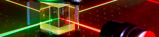
- LIGHT PROPERTIES CONTROL AT THE NANO-SCALE
- THREE ADVANCED MICROSCOPY APPROACHES
- FROM LIGHT FIELD STUDIES TO MICRO-OPTICS FABRICATION, TOWARDS THE MINIATURIZATION OF OPTICAL SYSTEMS
- QUANTUM IMAGING: FROM QUANTUM ILLUMINATION TO QUANTUM HYPOTHESIS TESTING
1. LIGHT PROPERTIES CONTROL AT THE NANO-SCALE
Plasmonic, alloyed and hybrid metasurfaces
Prof. Olivier J.F. Martin
Professor
Swiss Federal Institute of Technology, Lausanne (EPFL), CH
Bibliography
Olivier J.F. Martin is Professor of Nanophotonics and Optical Signal Processing at the Swiss Federal Institute of Technology, Lausanne (EPFL), where he is head of the Nanophotonics and Metrology Laboratory and Director of the Microengineering Section. He conducts a comprehensive research that combines modelling with nanofabrication and experiments on plasmonic systems, with applications in nonlinear optics, biosensing, security features and optical manipulations at the nanoscale. Dr. Martin has authored about 300 perr-reviewed publications and holds a handful of patents and invention disclosures. In 2005 he introduced the concept of an optical antenna, which is now widespread in plasmonic; in 2016 he received an ERC Advanced Grant on the utilization of plasmonic forces to fabricate nanosystems. Between 2016 and 2020, he served as director of the EPFL Microengineering Section and conducted with his colleagues major curricular reforms. He is associate editor to Advanced Photonics and Frontiers in Physics.
ABSTRACT: After a brief introduction to plasmonics, the optics of metal nanostructures, I will describe different technologies used for their fabrication with well-controlled features down to about 10 nm. Only a few plasmonic metals, such as gold, silver or aluminum, produce strong optical resonances, thus limiting the spectral range where plasmonics can be used. To extend that range, we recently developed a technology for the fabrication of Au-Ag alloyed nanostructures with well-controlled shapes that can be combined into metasurfaces to produce lenses or holograms. The working principle of these metasurfaces consists in engineering the phase associated with light scattered from metallic nanostructures to mimic the effects of gratings, lenses or phase plates. This scattered light can also be understood in terms of multipoles interactions. In the final part of the presentation, I will show that the available multipoles are especially rich and interesting when hybrid nanostructures that combine a plasmonic metal with a dielectric are considered.
Rolled-up multilayer nanostructures and their applications (VIDEO)
Humeyra Caglayan is an associate professor of physics in the Faculty of Engineering and Natural Sciences at Tampere University and leads the Metaplasmonics group. She received her Ph.D. degree in Physics from Bilkent University, Turkey, in 2010, where she investigated the novel electromagnetic phenomena in metamaterials and plasmonic structures. After her PhD studies, she worked as a postdoctoral scholar in Prof. Nader Engheta’s group at the University of Pennsylvania. Her group (Metaplasmonics) focuses on engineering the fundamental interaction between light and matter at the nanometer scale for plasmonic and metamaterial-based devices. She is an H2020 ERC Starting Grant holder (2019-2023).
ABSTRACT: Planar nanostructures, both dielectric and plasmonic, provide exceptional light manipulation at the subwavelength scale. Here, we introduce an out-of-plane nanohole-based nanostructure design with the implementation of a unique self-rolling technique. The photoresist-based technique enables the fabrication of nanostructures formed by nanohole arrays on gold (Au) and silicon dioxide rolled-up microtubes. The curved nature of the tube allows the fabrication of an out-of-plane nanostructure that can effectively control the wavefront compared to the common planar counterparts. This effect is verified by the spectral measurements of the fabricated samples.Besides, we implement a thin film self-rolling technique to fabricate SiO2/Au based three-dimensional metamaterials at visible wavelengths. Moreover, we investigate theoretically the potential of these structures for supporting an index zero waveguide mode inside the core of the RUTs. Our platform provides opportunities to integrate quantum emitters and 2D materials within index-zero structures with potential applications in quantum nanophotonics.
[1] M. Habib. et al., “Wavefront Control with Nanohole Array-Based Out-of-Plane Metasurfaces” ACS (2021). [2] D. Briukhanova, et al., “Low loss fishnet metamaterial via self-rolled nanotechnology” APL (2021). [3] I. Issah, M. Habib and H. Caglayan, “Long-range qubit entanglement via rolled-up zero-index waveguide” Nanophotonics (2021).
Physics, applications and integration of metasurfaces (VIDEO)
Patrice Genevet obtained his PhD at University of Nice Sophia Antipolis in France on localized spatial solitons in semiconductor lasers and amplifiers. After his PhD, he did five years of postdoctorate fellowship (2009-2014) in the Capasso group (SEAS, Harvard University) in collaboration with Prof. Scully (Texas A&M University). In 2014, he obtained the position of senior research scientist at A*STAR, Singapore. In 2015, He joined CNRS as ‘Chargé de Recherche de Première classe’. He is the recipient of the ERC starting Grant 2015 on Functional flat optical components and applications, the ERC Proof of Concept 2019 and the Aimé-Cotton Price 2017 from the French Optical Society. P. Genevet research activities concern the development of optical metasurfaces for sensing, imaging, and LiDAR applications. He authored 96 peer-review manuscripts and 6 international patents (H-factor 44, Citations ~20 000).
ABSTRACT: Metasurfaces are artificial optical interfaces designed to control the phase, the amplitude and the polarization of an optical wavefront. These optical surfaces rely on the coherent scattering of light by a sizable distribution of nanoscatterers of various shapes and material compositions. Metasurfaces hold great potential for on-chip integration of photonic components, significantly promoting the development of miniaturized optoelectronic systems. In this presentation, i will discuss on the physics of Metasurfaces and review some of our group results on Metasurfaces integration in VCSEL, and LiDARs. I will conclude this talk with perspective thoughts related to the developments in topological and tunable nanophotonics and applications.
[1] Q. Song, et al., Science Advances 7, no. 5, (2021) [2] Y Xie, et al., Nature nanotechnology (2020) [3] M. Elsawy, et al., Laser & Photonics Review, (2020) [4] Q. Song, et al., Nature Comms., 11, 2651 (2020) [5] I. Kim, R.J. Martins et al., Nature Nanotech. 16, (2021) [6] T Stolt, et al., Physical Rev. Lett. 126, 033901, (2021) [7] R Colom et al., (2022)
2. THREE ADVANCED MICROSCOPY APPROACHES
All-optical Super-resolution Imaging of Molecules in Their Nanoscale Cellular Context (VIDEO)
Joerg Bewersdorf is the Harvey and Kate Cushing Professor of Cell Biology and Professor of Biomedical Engineering and of Physics at Yale University. He received his Master’s degree (Dipl. Phys., 1998) and his doctoral degree in physics (Dr. rer. nat., 2002) training with Dr. Stefan W. Hell at the Max Planck Institute for Biophysical Chemistry in Goettingen, Germany. After 4 years at The Jackson Laboratory in Bar Harbor, Maine, he relocated his research group to Yale University in 2009. An optical physicist/biophysicist by training, Dr. Bewersdorf has been a long-time contributor to the field of super-resolution light microscopy development and the application of these techniques to cell biological questions.
ABSTRACT: Super-resolution optical microscopy has become a powerful tool to study the nanoscale spatial distribution of proteins of interest in cells over the last years. Imaging these distributions in the context of other proteins or the general cellular context is, however, still challenging. I will present recent developments of our lab which facilitate access to super-resolution microscopy for a broader community: A new fluorogenic DNA-PAINT probe enables fast, high-quality, 3D whole-cell imaging without the need for optical sectioning, adding a versatile and easily accessible tool to the toolbox of single-molecule super-resolution probes [1]. Labeling proteins and other cellular components in bulk in our recently developed pan-Expansion Microscopy method provides ultrastructural context to the nanoscale organization of proteins, replacing complex correlative light/electron microscopy by an all-optical approach to imaging cells and brain tissue sections [2, 3].
[1] Chung, K.K.H., et al. “Fluorogenic DNA-PAINT for faster, low-background super-resolution imaging”. Nat Methods (2022).
[2] M’Saad, O., Bewersdorf, J. “Light microscopy of proteins in their ultrastructural context”. Nat Commun 11, 3850 (2020).
[3] M’Saad, O., et al. “All-optical visualization of specific molecules in the ultrastructural context of brain tissue”. bioRxiv (2022).
Correlative imaging using ions and single molecules (VIDEO)
Aleksandra Radenovic is a full professor of biological engineering at the École Polytechnique Fédérale de Lausanne (EPFL) and head of the Laboratory of Nanoscale Biology. Her lab works in the research field that can be termed single-molecule biophysics. She has received her Ph.D. in Biophysics from the University of Lausanne (Switzerland.) in 2003 and a Msc. in Physics from the University of Zagreb (Croatia) in 2000. In 2010. she received a European Research Council (ERC) Starting Grant in 2010 and SNF Backup scheme Consolidator Grant (2015). She is also the recipient of the CCMX materials challenge award in 2016 and the Advanced ERC (2020) grant. She develops techniques and methodologies based on optical imaging, bio-sensing and single-molecule manipulation with the aim to monitor the behavior of individual biological molecules and complexes in vitro and in live cells.
ABSTRACT: From the plethora of correlative imaging modalities, SR techniques were most frequently combined with electron microscopy to provide protein-ultrastructure relationships at nanometer-scale resolution. At the other forefront of methods development, scanning probe microscopy techniques aim to capture nanoscale topographical dynamic changes of cells under physiological conditions. To avoid membrane deformation and to provide a method that could unlock long-term monitoring of the biological processes, we recently implemented SICM. The method currently experiences vast leaps in performance due to instrument developments and the ability to fabricate capillaries below tens of nanometers reliably. In contrast to AFM, SICM is truly non-contact, and represents the soft cell surface much more faithfully. In addition to providing accurate topographic imaging with nanometer resolution, SICM can be used to measure membrane stiffness surface charges and allows local delivery of material (e.g. fluorescent probes). The use of self-blinking dyes in SR microscopy permitted imaging conditions such as low laser excitation intensities and negligible bleaching that are ideal for live-cell imaging. In addition, the high SNR and photophysical properties of self-blinking dyes allow us to extend multiplane cross-correlation analysis to the 4th order using 8-plane volumetric imaging, achieving up to 29 planes. Finally, with a combined SICM-SR setup we demonstrate long-term correlated live-cell imaging.
Beyond CLEM: Multiscale and 3D Volume Imaging across Light, Electron and X-Ray Microscopy (VIDEO)
Francesco Biancardi got his Master degree in Electronics Engineering and Executive MBA at Politecnico di Milano, passionate for Scientific Research and Technology, he worked as Specialist, Sales Account Manager and Business Development in different fields including Light, Electron, X-Ray, Atomic Force microscopy, interferometry, Tribology. Since 2017 he supports customers in the acquisition and implementation of Zeiss Research Microscopy Solutions, in particular SEMs, Xradia and Correlative Workflows
ABSTRACT: How to match multiple perspectives of a sample across scales, different image modalities and analytical approaches and finally answer the Scientific Questions? Whether it is Confocal, Super Resolution or Electron microscopy, each imaging system has its strengths and gives access to information that was hidden before. Combining techniques with new technology solutions opens the way to the next level of imaging and 3D Volume reconstruction in Life Sciences
3. FROM LIGHT FIELD STUDIES TO MICRO-OPTICS FABRICATION, TOWARDS THE MINIATURIZATION OF OPTICAL SYSTEMS
Microscopes on chip for cellular imaging
Andrea Bassi received his Ph.D. in Physics at Politecnico di Milano in 2006. He conducted research at Beckman Laser Institute, University of California (Irvine) in 2005/2006 as Research Specialist, at Politecnico di Milano from 2009 to 2014 as Researcher, at Max Planck Institute of Molecular Cell Biology and Genetics (Dresden) in 2013/2014 as Marie Curie Fellow. Currently he is an Associate Professor at the Department of Physics of Politecnico di Milano. His scientific interests include optical tomography and microscopy for biological and preclinical applications.
ABSTRACT: Opto-fluidic technologies integrate multiple fluidics and photonics devices in a single chip. Recently these technologies have shown their potential in the field of optical microscopy [1] for biological imaging. The talk will discuss the design and applications of miniaturized optofluidic devices for fluorescence optical microscopy. First, an optofluidic chip that incorporates light-sheet illumination and automatic sample delivery will be illustrated. This device upgrades a standard inverted microscope to automatic, three-dimensional, Light Sheet Fluorescence Microscope. Then a miniaturized device, based on integrated waveguides will be shown, as a new source for structured illumination microscopy. Finally, the combination of these two technologies will be described to demonstrate automatic imaging of cells at enhanced resolution. Example applications will be discussed, together with the technological solutions for automatic sample alignment, including automatic imaging of tumour spheroids, Drosophila embryos, and high-resolution imaging of single cells [2].
[1] Paiè, P., Martínez Vázquez, R., Osellame, R., Bragheri, F., and Bassi, A. “Microfluidic based optical microscopes on Chip” Cytometry Part A, 93, 987-996 (2018). [2] Sala, F., Castriotta, M., Paiè, P., Farina, A., D’Annunzio, S., Zippo, A., Osellame R., Bragheri F., Bassi, A. (2020). High-throughput 3D imaging of single cells with light-sheet fluorescence microscopy on chip. Biomedical optics express, 11(8), 4397-4407.
High-resolution 3D printing of novel on-fiber optical structures (VIDEO)
Carlo Liberale is heading the Vibrational Imaging (VIBRA) Lab at the King Abdullah University of Science and Technology (KAUST, Saudi Arabia). He received his Ph.D. in Electrical Engineering from the University of Pavia (Italy). His research group focuses on developing and applying label-free chemical microscopy techniques based on vibrational spectroscopy. Additionally, his lab uses high-resolution 3D printing based on two-photon lithography to miniaturize complex optical systems. Using this approach, he has pioneered the fabrication of novel micro-optical components directly integrated at the end-face of optical fibers, for applications including beam shaping, optical tweezers, and micro-endoscopy. This research activity takes advantage of an integrated approach that combines design, micro/nanofabrication, and optical techniques. He has authored more than 70 publications and height patent applications. He is a Senior Member of the Institute of Electrical and Electronics Engineers (IEEE) and the Optical Society of America (OSA).
ABSTRACT: We leverage the potential of 3D micro-printing to develop novel micro-optic devices for bio-photonic applications. This cutting-edge technology provides unprecedented three-dimensional micro-fabrication capabilities, and aligns with the increasing demand for the miniaturization of optical probes and setups towards compact, portable, less-invasive, and point-of-care smart devices. Within this research line, my group has demonstrated new concepts on how to control the light output from optical fibers, often enabling previously unattainable on-fiber functionalities for the first time. In this seminar, I will present our work on designing and fabricating novel miniaturized 3D optical systems for different applications, including beam shaping, polarization control, micro-endoscopy, orbital angular momentum generation, and optical tweezers.
[1] I. Reddy, A. Bertoncini, C. Liberale, 3D-printed fiber-based zeroth- and high-order Bessel beam generator, Optica 9(6), 645-651 (2022) [2] S. Sivankutty, A. Bertoncini, V. Tsvirkun, N. G. Kumar, G. Brévalle, G. Bouwmans, E. R. Andresen, C. Liberale, H. Rigneault, Miniature 120-beam coherent combiner with 3D-printed optics for multicore fiber-based endoscopy, Opt. Lett. 46, 4968-4971(2021). [3] A. Bertoncini, C. Liberale, 3D printed waveguides based on photonic crystal fiber designs for complex fiber-end photonic devices, Optica 7(11), 1487-1494 (2020). [4] A. Antonini, A. Sattin, M. Moroni, S. Bovetti, C. Moretti, F. Succol, A. Forli, D. Vecchia, V.P. Rajamanickam, A. Bertoncini, S. Panzeri, C. Liberale, T. Fellin “Extended field-of-view ultrathin microendoscopes for high-resolution two-photon imaging with minimal invasiveness,” eLife 9, e58882 (2020)
Spin-orbit shaping of light with/from topological defects (VIDEO)
Etienne Brasselet obtained a joint PhD in 2001 from University Paris-Sud, Orsay, in France and Laval University, Quebec, in Canada on the optical manipulation of liquid crystals. After his PhD, he worked as a postdoctorate fellow for five years, exploring nonlinear optics of doped polymers (Ecole Normale Supérieure de Cachan, France, 2001-03), nonlinear dynamics (Laval University, Quebec, Canada, 2003-04) and the physics of liquid crystals (Ecole Normale Supérieure de Lyon, France, 2004-06). In 2007, he was recruited as CNRS researcher at Laboratoire Ondes et Matière d’Aquitaine where he is now leading a research group. He is the recipient of the Ancel Prize 2019 from the French Physical Society. His research interests mainly focus on the interaction of electromagnetic and acoustic waves with soft or solid-state matter whenever structured fields meet structured matter, which involves the physics of singularities and topological defects.
ABSTRACT: Since two decades, substantial research and technological efforts have been made to develop optical elements enabling the versatile manipulation of light fields via geometrical principles, which manifest as the so-called geometric phase that finds its root in the optical spin-orbit interaction. Several assets make this approach especially attractive. Indeed, being geometrical by nature, such a beam shaping approach is particularly suitable to process polychromatic light fields. Also, coupling the polarization state of light to the spatial degrees of freedom allow considering novel perspectives in optical information, optical imaging, optomechanics, optical material processing, and optical sensing. In addition to current technologies that make it possible to fabricate flat optics with versatile beam shaping capabilities, we will examine how the self-organization of liquid crystals enables nature-assisted approaches to advanced topological light shaping that open up new applications.
The BrightEyes project: towards a new generation of laser-scanning microscopy for imaging, tracking and spectroscopy (VIDEO)
Giuseppe Vicidomini studied computer science at the University of Genoa and received his Diploma cum laude in 2003. From 2003 to 2007, he worked at the LAMBS (University of Genoa), receiving his PhD in image processing and analysis for fluorescence microscopy. From 2008 to 2011, he worked as a post-doctoral fellow at the MPI, where he developed a new method which allows stimulated emission depletion (STED) microscopy to achieve tens of nanometres spatial resolution with a substantial reduction of the dose of light requested. Since May 2011, he have worked at the IIT, where in May 2016, he became the principal investigator of the Molecular Microscopy and Spectroscopy research line. While constantly working on STED microscopy and its combination with fluorescence correlation spectroscopy (STED-FCS), he also started developing novel single-photon-avalanche diode (SPAD) arrays for fluorescence microscopy. This research has recently evolved in the introduction of a new paradigm which leverages the single-photon information to implement a high-resolution and high-informative laser-scanning microscope for imaging, spectroscopy and particle tracking. He is a co-founder and scientific advisor of the Genoa Instruments spinoff company.
ABSTRACT: Fluorescence laser-scanning microscopy (LSM) is one of the most popular microscopy architectures. This success comes from its versatility, ability to work across different spatial and temporal scales, and access to many samples’ information. However, in a conventional LSM system, how it records the fluorescent signal induces a substantial loss of information. Our group has introduced a new detector (i.e., a single-photon avalanche diode (SPAD) array detector) specifically designed to provide access to spatial and temporal information otherwise discarded by a typical single-element detector. The aim of the BrightEyes project is to leverage this new spatial and temporal information to enhance all significant abilities of fluorescence LSM further and to open new exciting perspectives for imaging, spectroscopy, and particle/molecule tracking. In this seminar, I will briefly overview the BrightEyes project’s results in developing a super-resolution fluorescence lifetime imaging system [1] and a high-informative fluorescence fluctuation spectroscopy method [2]. Later, I will present a new implementation of single-particle/molecule tracking and its perspective for investigating dynamics in thick multi-cellular samples.
[1] M. Castello, G. Tortarolo, M. Buttafava, T. Deguchi, F. Villa, S. Koho, L. Pesce, M. Oneto, S. Pelicci, L. Lanzanò, P. Bianchini, C. J. Sheppard, A. Diaspro, A. Tosi, and G. Vicidomini, “A robust and versatile platform for image scanning microscopy enabling super-resolution FLIM,” Nat. Methods, 16(2):175-178, 2019. [2] E. Slenders, M. Castello, M. Buttafava, F. Villa, A. Tosi, Lanzanò, S. V. Koho, and G. Vicidomini, “Confocal-based fluorescence fluctuation spectroscopy with a SPAD array detector,” Light Sci. Apll., 10:31, 2021.
4. QUANTUM IMAGING: FROM QUANTUM ILLUMINATION TO QUANTUM HYPOTHESIS TESTING
Hong-Ou-Mandel microscopy and fluorescence lifetime imaging (VIDEO)
Daniele Faccio is a Royal Academy Chair in Emerging Technologies, Fellow of the Royal Society of Edinburgh and Cavaliere dell’Ordine della Stella d’Italia (Knight of the Order of the Star of Italy). He joined the University of Glasgow in 2017 as Professor in Quantum Technologies where he leads the Extreme-Light group and is Director of Research for the School of Physics and Astronomy. He is also adjunct professor at the University of Arizona, Tucson (USA) and fellow of the Optical Society of America. Previously he was at Heriot-Watt University and University of Insubria (Italy). He has been visiting scientist at MIT (USA), Marie-Curie fellow at ICFO, Barcelona (Spain) and EU-ERC fellow 2012-2017. He was awarded the Philip Leverhulme Prize in Physics in 2015, the Royal Society of Edinburgh Senior Public Engagement medal and the Royal Society Wolfson Merit Award in 2017. He worked in the optical telecommunications industry for four years before obtaining his PhD in Physics in 2007 at the University of Nice-Sophia Antipolis (France). His research, funded by the UK research council EPSRC, DSTL, The Leverhulme Trust, the EU Quantum Flagship program and the Royal Academy of Engineering, focuses on the physics of light, on how we harness light to answer fundamental questions and on how we harness light to improve society.
ABSTRACT: Hong-Ou-Mandel or two-photon interference is a widely used effect in quantum optics with applications including sensing path length changes all the way down to the nanometre scale, filtering of quantum states and teleportation and also forms the basis of quantum computing based on Boson sampling. We will report on recent work in which we have extended the use of HOM sensing to the imaging domain for microscopy applications [1] and will briefly overview some of the main constraints such as the biphoton mode-matching conditions between the quantum light and the camera pixels that need to be considered. We will then present a development (unpublished work) in which we use HOM interference applied to the case of fluorescence lifetime imaging, a widespread microscopy technique. This FLHOM technique provides access to very short fluorescence lifetimes, i.e. lifetime resolution in the picosecond range that is therefore 100x better that what can be achieved with standard TCSPC approaches. We demonstrate the advantage of this for the case of viscosity measurements in which we measure the fluorescence lifetime of “rotor” molecules, thus providing a new approach to nanorheology with applications for intracellular in-vivo measurements that are not currently otherwise possible.
[1] Quantum microscopy based on Hong–Ou–Mandel interference
Scanning Fluorescence Microscope with Entangled Two-Photon Excitation
Oleg Varnavski received Master of Science from the Moscow Institute of Physics and Technology in 1974 and his Ph.D. from the P.N. Lebedev Physics Institute of Russian Academy of Sciences in 1984. He received Alexander von Humboldt Fellowship (Germany) and worked at Stuttgart University in 1986 and 1990. Dr. Varnavski joined the research group of Professor Goodson at Wayne State University (Detroit, USA) in 1999. He is currently a research scientist at Chemistry Department of the University of Michigan (Ann Arbor, USA). His research interests include ultrafast photodynamics in organic materials, energy transfer processes in complex macromolecules (coherent and incoherent), in nanoscale metal and semiconductor systems, nonlinear optics and spectroscopy, quantum optics, specifically quantum entangled photons for spectroscopy and imaging.
[1] Eshun, A.; Varnavski, O.; Villabona-Monsalve, J.P.; Burdick, R.K.; Goodson III, T. Entangled Photon Spectroscopy Acc. Chem. Res. 2022, 55, 991−1003 [2] Varnavski, O. ; Goodson III, T. Two-Photon Fluorescence Microscopy at Extremely Low Excitation Intensity: The Power of Quantum Correlations. J. Am. Chem. Soc. 2020, 142, 12966–12975. [3] Varnavski, O.; Gunthardt, C.; Rehman, A.; Luker, G.D.; Goodson III, T. Quantum Light-Enhanced Two-Photon Imaging of Breast Cancer Cells J. Phys. Chem. Lett. 2022, 13, 2772−2781.
Application in Quantum Hypothesis Testing: quantum reading and pattern recognition (VIDEO)
Giuseppe Ortolano is a researcher at the Quantum Optics group at the INRiM (Istituto Nazionale per la Ricerca Metrologica) in Torino, with a research grant on the study of quantum techniques to improve classical schemes. He got his Master degree in “Physics of complex Systems” from the University of Turin in 2019. He did his PhD in Physics at Politecnico di Torino. His main research interest is quantum information and quantum optics. In his research work he have treated different problems in quantum metrology and quantum hypothesis testing, both theoretically and experimentally.
ABSTRACT: The aim of Quantum Hypothesis Testing is to reach an advantage over all classical strategies. In the quantum reading (QR) protocol [1] quantum resources are used to enhance the readout of classical data stored in optical supports. In the single-cell scenario a bit of information is stored in one of two possible values of transmittance (or reflectance). Practically, an advantage is achieved using quantum-correlated photon sources and photon-counting measurements [2,3]. A significant experimental sensing advantage can be obtained with this strategy even in presence of not negligible optical losses, showing the robustness of the proposal.
The single-cell quantum sensing advantage is preserved in higher level tasks, like pattern recognition (PR). In PR (e.g. handwritten digit classification), the relevant information is spread non-trivially over complete images. We experimentally show how the advantage in sensing is preserved in PR, using different classification algorithms. Our findings highlight the scalability of the quantum enhancement in sensing, in the context of QR, to more complex scenarios, an important feature in view of practical applications.
[1] S. Pirandola, Quantum reading of a classical digital memory, Phys. Rev. Lett. 106, 090504 (2011). [2] G. Ortolano, E. Losero, S. Pirandola, M. Genovese, and I. Ruo-Berchera, Experimental quantum reading with photon counting, Sci. Adv. 7, eabc7796 (2021). [3] G. Ortolano, I. Ruo-Berchera, Quantum readout of imperfect classical data, Sensors 22, 2266 (2022)
Videos:
T1 Speakers
T1 Events